| In order to understand the advantages of
harmonic imaging, we have to look at two acoustic phenomena: grating
lobes and harmonics.
Grating lobes disturb the image
Due to the physical construction of an ultrasound transducer, secondary
beams called grating lobes split off at an angle to the main ultrasound
beam. When grating lobes are reflected from other structures or fatty
tissue, they produce unwanted effects on the ultrasound image as they
return to the transducer. The problem is often seen in low-intensity
areas of the image. Breast scanning and prostate scanning are examples
of applications where grating lobes may reduce image clarity.
Harmonics offer a different
kind of signal
Conventional ultrasound imaging sends out a fundamental beam and
receives essentially the same frequency range back as an echo. However,
the sound wave becomes distorted as the tissue expands and compresses in
response to the wave. When a certain energy level is reached, this
distortion results in the generation of additional frequencies, called
harmonics, that are two, three or more times the emitted frequency. The
harmonic frequencies return to the transducer together with the
fundamental frequency.
Harmonics arise only in the fundamental frequency, not
in grating lobes, because the energy level of the grating lobes is not
high enough. The second harmonic (twice the frequency of the
fundamental) is the frequency used for harmonic imaging.
Using harmonics to reduce
the effect of grating lobes
Although the harmonic signal is weaker than the fundamental beam, it
better retains its purity by only having to travel one way, from within
the tissue (where it is generated) outwards to the receiver. The
accumulation of noise and clutter is significantly reduced, because the
harmonic signal only has to travel from the tissue back to the
transducer.
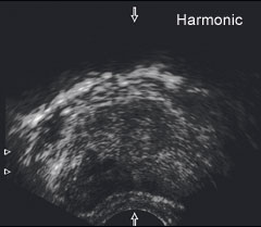
By electronically filtering out the fundamental
frequency and its grating lobes, the harmonic frequency can be isolated
and can be used to create an alternative ultrasound image. Compared with
standard mode, the harmonic image will typically show enhanced contrast
and gray tone differentiation.
Most effective in the
mid-range
Because harmonics are generated within the tissue itself, a certain
distance from the sender is required in order for the harmonic wave to
build up. Near-field imaging does not permit this. Conversely, signal
intensity and sensitivity are lost in the far field. Therefore, harmonic
imaging is most applicable when scanning structures in the middle range.
Transducers with True Echo
Harmonics
Harmonic imaging is available with a number of B-K Medical transducers.
Click here for
an overview. |
Grating lobes occur because secondary beams split off
from the main ultrasound signal, due to certain physical characteristics
of the ultrasound system and transducer.
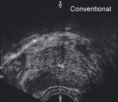
Verified malignancy in the right posterior peripheral
zone of the prostate, seen in standard imaging mode.
|
