Home Enquiry Form Quotation Request Forms Brochure Request Form What's New Product Lists Product User Manuals Product Technical Notes Ultrasound Equipment Ultrasound Transducers Seeding/Biopsy Steppers Disposable Grids Disposable Drapes Endocavity Balloons Grid Adapters PAL Pro Stirrups OR Tables / Trolleys Source Measurement Electrometers Equipment Services Suggestions FAQ Search Useful Web Sites Training Courses Calibrations and Upgrades Intravascular Brachy LDR Calibrations Prostate Brachytherapy Prostate Biopsy 3D Prostate Mapping Prostate Cancer Prostate Seed Implant Mammography/Diagnostics Site Map External Beam/IMRT/CT Exradin Ion Chambers Prostate Scanning SeeDs and Strands Prostate Seeding Needles Stabilisation Needles LDR,HDR,PDR Dose Planning Ir-192 Seeds in Ribbon HDR Prostate Brachy HDR/LDR Templates AUTIMO Beam Systems Alloys and Alloy Melters Slideshow Bibliography Our Company Urology Biopsy Biopsy Guns/Needles Needles/Guns/Applications Clinical Oncology Needles Haematology Biopsy Interventional Radiology Urology Needles/Guns Breast Biopsy/Localisation Gynae Biopsy / Aspiration Skin Biopsy Punch Anaesthesia Needles Antenatal Diagnostic Needle Guides Syringe Holders Biopsy Grids Biopsy Steppers Infection Control-MultiMAT Bone Marrow Biopsy Vertebroplasty Laparoscopy Needles Laparoscopy Support Temno Adjustable Coaxial MRI Biopsy/Localisation Percutaneous Injection-PIA Portable Bladder Scanner Cryotherapy Brachytherapy Template Guided Biopsy Volume Studies Gold Fiduciary Markers Fiducial Marker Placement Micro-Touch Micro-Touch LP NEW EX3 Stepper Classic Stepper Multi-Purpose Workstation Stabilizers Steppers Brachy/Biopsy Accessories Radiotherapy Immobilization Rehabilitation Fixation
 

 Ultrasound Transducers
Home
Enquiry Form
Quotation Request Forms
Brochure Request Form
What's New
Product Lists
Product User Manuals
Product Technical Notes
Ultrasound Equipment
Ultrasound Transducers
Seeding/Biopsy Steppers
Disposable Grids
Disposable Drapes
Endocavity Balloons
Grid Adapters
PAL Pro Stirrups
OR Tables / Trolleys
Source Measurement
Electrometers
Equipment
Services
Suggestions
FAQ
Search
Useful Web Sites
Training Courses
Calibrations and Upgrades
Intravascular Brachy
LDR Calibrations
Prostate Brachytherapy
Prostate Biopsy
3D Prostate Mapping
Prostate Cancer
Prostate Seed Implant
Mammography/Diagnostics
Site Map
External Beam/IMRT/CT
Exradin Ion Chambers
Prostate Scanning
SeeDs and Strands
Prostate Seeding Needles
Stabilisation Needles
LDR,HDR,PDR Dose Planning
Ir-192 Seeds in Ribbon
HDR Prostate Brachy
HDR/LDR Templates
AUTIMO Beam Systems
Alloys and Alloy Melters
Slideshow
Bibliography
Our Company
Urology Biopsy
Biopsy Guns/Needles
Needles/Guns/Applications
Clinical Oncology Needles
Haematology Biopsy
Interventional Radiology
Urology Needles/Guns
Breast Biopsy/Localisation
Gynae Biopsy / Aspiration
Skin Biopsy Punch
Anaesthesia Needles
Antenatal Diagnostic
Needle Guides
Syringe Holders
Biopsy Grids
Biopsy Steppers
Infection Control-MultiMAT
Bone Marrow Biopsy
Vertebroplasty
Laparoscopy Needles
Laparoscopy Support
Temno Adjustable Coaxial
MRI Biopsy/Localisation
Percutaneous Injection-PIA
Portable Bladder Scanner
Cryotherapy
Brachytherapy
Template Guided Biopsy
Volume Studies
Gold Fiduciary Markers
Fiducial Marker Placement
Micro-Touch
Micro-Touch LP
NEW EX3 Stepper
Classic Stepper
Multi-Purpose Workstation
Stabilizers
Steppers
Brachy/Biopsy Accessories
Radiotherapy Immobilization
Rehabilitation Fixation


Ultrasound Transducers

For general transducer application information please go to TRANSDUCER INFORMATION

Compatibilities for urology specific transducers
 
Compatibilities
8803
Prostate
Kidney
*
Testis
Penile
Pro Focus UltraView
Flex Focus
Flex Focus Solo
Falcon Premium
Pro Focus Blue(B)
Pro Focus Green(C)
Pro Focus Yellow(CX)
Contrast **
Tracked 3D
Doppler
HistoScanning™
Biopsy guide
 

Pro Focus UltraView + 8818, 8808 or 8667
Flex Focus + 8818, 8808e or 8667
Flex Focus Solo + 8808e or 8667


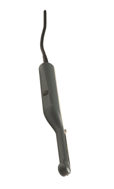
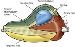
Triplane – all prostate zones with one transducer
  • unsurpassed images in 3 visionary planes
  • switch between prostate zones at the touch of a button
  • increase diagnostic value with 3D, Contrast and Doppler
Easy and comfortable to use
  • take confident apical biopsies with endfire array
  • biopsy the peripheral, transition and central zones with simultaneous biplane
  • one-time insertion and minimal manipulation using disposable dual guide
Read more about our sterile single-use needle guides here.
8818 Applications
  • Transrectal prostate scanning
  • Transrectal puncture and biopsy
  • Transperineal puncture and biopsy
  • Transvaginal scanning
  • Spectral and CFM Doppler examinations
  • Tissue harmonic imaging
  • Contrast imaging*
 
 
8818 Specifications
Frequency range 4 - 12 MHz
Contact surface (acoustic) 34.4 x 5.5 mm
Focal range 3 - 60 mm
Scanning modes B, M,BCFM, Doppler, Contrast*, Tissue Harmonic
Frame Rate 60 Hz
Image field (expanded) Triplane / 140°
Dimensions 36 x 39 x 323 mm
Weight 230 g
Disinfection Immersion,
Steris system 1
®, Sterrad®
50,100S and 200
 
 
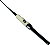
Simultaneous biplane: the gold standard
  • for orientation and correct biopsy spacing
  • real-time images of both the sagittal and transverse planes
  • confidently take core biopsies
Multifrequency simultaneous biplane probe
  • high frequency for excellent near field scanning
  • excellent power doppler
3D and harmonic imaging for easier identification of lesions
  • use 3D as a diagnostic tool
  • visualization of lesions in 3 planes appears to allow improved assessment of capsular disruption
Dedicated biopsy guides
  • easy cleaning and contamination prevention
  • the needle path is angled optimally through the channel bracket instead of running along the shaft of the transducer
Read more about our sterile single-use needle guides here.
 
 
8808 Specifications
Frequency range 6 - 10 MHz
Contact surface (acoustic) 5 x 19.6 mm
Scanning Modes B, M, Doppler, BCFM, Tissue Harmonic
Focal range 5 - 50 mm
Sector Angle biplane / 127°
Disinfection Immersion, STERIS SYSTEM 1®
Dimensions 120 x 21 mm
Weight


Endfire prostate imaging
  • wide endfire image plane enables you to locate a lesion and then rotate the plane around a central axis.

A balance between resolution and penetration

  • multifrequency capability enables reduction of frequency to 6 Mhz without removing the transducer
Convenient puncture
  • the needle's path starts at the tip of the probe
  • monitor the needle from the start of the puncture to the actual biopsy site
8667 Applications
  • Endorectal scanning
  • Interventional procedures
  • Spectral, CFM and Power Doppler examinations
  • Tissue harmonic imaging
 
 
8667 Specifications
Frequency range 6 - 10 MHz
Contact surface (acoustic) 50 mm2
Focal range 5 - 50 mm
Scanning modes B, M, BCFM, Doppler, Tissue Harmonic, Power Doppler
Disinfection Immersion, STERIS SYSTEM 1®
Dimensions 300 x 36 mm
Weight 260 g


Image guided prostate therapy
  • No gland too large — sagittal scanning of any size prostate from base to apex
  • Resolute, clear and detailed image, for accurate volume studies and source dose planning
  • Customizable sagittal grids and preferences for brachytherapy
  • Clear visualization of seminal vesicles
  • Clear view of needle placement                                           

 Pelvic Floor Scanning

  • Best broad view of anterior and posterior compartments for functional and anatomical studies
  • Reproducible 3D studies with external mover
  • Detailed high-resolution biplane with 6.5 cm. linear and convex views
     

    * In the USA, contrast-enhanced ultrasound has not been market cleared by the FDA, with the exception of only select cardiac imaging applications.


8848 Specifications
Frequency range 5-12 MHz
Focal range
(typical)
3-60 mm
Frame rate >150
Disinfection Immersion,
Steris system 1
®, Sterrad®
50,100S and 200
Scanning modes B,M, Doppler, BCFM, Tissue Harmonic Imaging
Contact surface (overall) Transverse:
127 mm2

Sagittal:
357 mm2
Image field
(expanded)
180˚(transverse)
Weight (approx.) 250 g


Ultrasound Transducer, SeeDOS Code 8658

Now replaced by the more advanced transducers shown above

 
Unique biplane imaging: Two planes are better than one
  • biplanar with multifrequence capabilities
  • long sagittal array for base to apex view in seed implantation procedures
  • easier prostate volume determination
Colour Doppler and transperineal facilities
  • excellent for all transperineal procedures including brachytherapy
  • transperineal needle guide
8658 Applications
  • Prostate brachytherapy
  • Transrectal scanning
  • Transperineal puncture
  • Transvaginal scanning
  • Intraoperative scanning
  • Cryosurgery
  • Spectral and CFM Doppler examinations
 
 
8658 specifications
Frequency range 5 - 7.5 MHz
Sector angle 110° for transverse, 60 mm for sagittal
Focal range 70 mm
Scanning modes B, M, BCFM, Doppler, Power Doppler
Disinfection Immersion, EO
Dimensions 340 x 32 mm
Weight 280 g


UltraView and 8823 with needle guide


Easy access to kidney diagnostics
  • superior image quality
  • minimizes patient discomfort
Exceptional image quality
  • clearly view structures using deep penetration at higher frequencies obtaining a high image resolution with coded excitation
  • measure renal blood flow with superb spectral Doppler
  • visualize anatomic variations and residual tumor after RF and cryoablation with contrast imaging
  • find kidney stones easily with harmonic imaging
Ideal for interventional procedures
  • single-use and reusable needle guides for convenient interventional procedures
8823 Applications
  • Kidney
  • Bladder
  • Pediatric
  • Difficult to access areas
 
 
8823 Specifications
Frequency range 1.8 - 6 MHz
Contact surface 31 x 12 mm
Penetration depth 26 cm
Scanning modes
B, M, Doppler, BCFM, Contrast, Tissue Harmonic
Disinfection
Immersion, sterile covers,STERIS
SYSTEM 1®, single-use and reusable puncture guides.
Dimensions 94 x 44 mm
Weight 150 g


* In the USA, contrast-enhanced ultrasound has not been market cleared by the FDA, with the exception of only select cardiac imaging applications.


The most advanced laparoscopic ultrasound transducer on the market
  • built in facilities for LUS-guided biopsies
  • RFA
  • ethanol or contrast agent injections
  • cryoablation
  • microwave ablation
  • flexible for difficult to reach areas or
  • rigid for manipulating structures
8666-RF Applications
  • Laparoscopic
  • Intraoperative
  • Radiofrequency tumor ablation (RFA)
  • Biopsy
  • Drainage

* In the USA, contrast-enhanced ultrasound has not been market cleared by the FDA, with the exception of only select cardiac imaging applications.


8666-RF Specifications
Frequency range 5 - 10 MHz
Focal range (typical) 5 - 95 mm
Contact surface 30 x 5 mm
Sector angle 36°
Scanning modes B, M, Doppler, BCFM,
Tissue Harmonic Imaging
and Contrast Imaging*
Disinfection Immersion, EO (ethylene oxide),
STERIS SYSTEM 1® or STERRAD®
Dimensions 302 x 178 mm
Weight 475 g


Deep penetration and high resolution
  • clearly visualize deep anatomical structures
  • coded excitation
Comfortable ergonomic design
  • a control button right on the handle
  • slim design and rounded handle enables lighter grip with minimum pressure
More diagnostic information
  • contrast imaging*
8820e Applications
  • Liver
  • Pancreas
  • Bladder
  • General abdominal
  • Obstetric scanning
  • Interventional procedures
 
 
8820e Specifications
Frequency range 2 -6 MHz
Contact surface (acoustic) 62.5 x 13 mm
Scanning modes B, M, Doppler, BCFM, Tissue Harmonic, and Contrast*
Focal range 12 - 200 mm
Disinfection Immersion
Dimensions 104 x 77 mm
Weight 180 g


* In the USA, contrast-enhanced ultrasound has not been market cleared by the FDA, with the exception of only select cardiac imaging applications.


Quality, versatility and convenience in one transducer

Small part, musculoskeletal and vascular scanning. The 8670 supports a number of ultrasound applications, such as small part, breast and orthopedics, rheumatology and sports medicine.

  • Easy switching between near and far views
  • Very high image detail
  • Excellent choice for penile Doppler and testis
  • Optimized for small part, musculoskeletal and vascular scanning
  • Easy-to-use puncture and biopsy guide
  • Small part
  • Breast
  • Testis
  • Penile Doppler
  • Musculoskeletal
  • Peripheral vascular
  • Interventional procedures
  • Contrast Imaging
 
 
8670 specifications
Frequency range 5 - 12 MHz
Contact surface 45 x 14 mm
Focal range 0 - 70 mm
Scanning modes B, M, BCFM, Doppler, Tissue Harmonic
Disinfection Immersion
Dimensions 91 x 52 x 21 mm
Weight 130 g


* In the USA, contrast-enhanced ultrasound has not been market cleared by the FDA, with the exception of only select cardiac imaging applications.


A clear choice for cost-effective imaging
  • very high frequencies
  • large footprint
  • Doppler sensitivity
  • True Echo Harmonics
Puncture guides for convenient biopsy
Puncture guides available have:
  • 30, 45 and 60 angles of insertion.
  • variable diameter, allowing you to choose the desired needle size.
Disposable needle guides and sterile transducer covers are also available.

Immediate lesion assessment

  • provides image clarity and contrast resolution
  • determine necessity for biopsy on the spot
  • perform aspiration or biopsy immediately
 
 
8811 Specifications
Frequency range 5 - 12 MHz
Focal range (typical) 2 - 55 mm
Acoustic contact 50 x 4 mm
Disinfection Immersion
or STERIS SYSTEM 1
Dimensions 105 x 64 x 22 mm
Weight 98 g
                                                                Prostate Scanning


Prostate anatomy
The prostate consists of five zones: three glandular (the peripheral, transition, and central zone) and two non-glandular (periurethral zone and fibromuscular stroma).

Peripheral
A healthy peripheral zone displays a homogeneous isoechoic ultrasound pattern. The “surgical capsule” that separates the peripheral zone from the transition zone appears as a distinct boundary in ultrasound.

Transition
The transition zone consists of two separate lobes that lie superior to the verumontanum, lateral to the proximal urethra, and posterior to the fibromuscular stroma.

Central
The central zone lies posterior to the urethra and superior to the verumontanum and surrounds the ejaculatory ducts. The central zone makes up 25% of the glandular tissue.

Anterior fibromuscular stroma
The anterior fibromuscular stroma forms the anterior surface of the gland. It may be up to 1cm thick, but thins out over the distal portion of the gland. The anterior fibromuscular stroma is thought to be a barrier to the spread of cancer.

Periurethral
The periurethral zone is a mid-line structure comprised of a cylindrical, internal smooth muscle sphinter that run from the base of the verumontanum to the back of the bladder neck. Its function may be to prevent semen from flowing backwards into the bladder.


Prostate Doppler
Colour Doppler is used as a tool for the detection of prostatis.

In prostate cancer diagnosis and follow-up after therapy, the use of color Doppler guides the biopsies of focal lesions and the detection of reoccurrence.


Renal ultrasound solution
UltraView + 8823 + 8666 + 8820e
or
Flex Focus + 8823 +8666

 
Kidney anatomy
Imaging the kidneys can be a challenge because of their depth. The kidneys are hidden behind dense muscles and the shadow of the ribs. Our transducers compensate for anatomic constraints and produce clear images with deep penetration during intercostal scanning.

Urologic ultrasound of the kidney
Ultrasonography for renal diagnostics is inexpensive, safe, and fast. Renal ultrasound provides information about the location, shape, size, and outer shell thickness of the kidney. Ultrasound is a tool to screen for kidney stones, cysts, and masses. Detailed information from a scan can be used to accurately choose the best course of therapy.

The Pro Focus UltraView, 8823 or 8820e, and laparoscopic 8666 provide a complete renal scanning package.

 
8823

8820e

8666
A powerful renal ultrasound solution

The UltraView, 8823 and laparoscopic 8666 provide a complete renal scanning package.

 
  • The 8823 allows you to clearly view structure in the kidney using deep penetration at higher frequencies obtaining a high image resolution with coded excitation. Spectral Doppler makes it is possible to measure renal blood flow. Find kidney stones easily using harmonic imaging. Designed with a rounded scanning surface, the 8823 makes it possible to shorten scan time and minimize patient discomfort with easy access between the ribs and better contact with the skin.
  • 8820e provides comfort and quality in abdominal ultrasound. The 8820e delivers the depth and sharpness needed for ultrasound images. The slim design and rounded handle of the 8820e enable a lighter grip with minimal pressure.
  • The 8666 is the most advance laparoscopic ultrasound transducer on the market. It has a versatile built-in biopsy guide for staging. The 8666 provides rigid and 4-way flexible tip to reach difficult to access areas. Laparoscopic ultrasound is less invasive than conventional surgery, and therefore decreases the risk of complications, especially in patients where major surgery is not indicated.
  • UltraView enables Contrast Enhanced Ultrasound*, which is helpful in the immediate assessment of residual tumour after RF and other ablative treatments.


    * In the USA, contrast-enhanced ultrasound has not been market cleared by the FDA, with the exception of only select cardiac imaging applications.


UltraView + 8670 or 8811

Flex Focus + 8670 or 8811

8670

8811


Testicular ultrasound
With our ultrasound equipment it is possible to:
• Examine a suspicious mass
• Monitor infection or inflammation
• Evaluate injuries
• Identify testicular torsion
• Check for vascularity

Varicoceles
The presence of varicoceles is a leading factor in the cause of male infertility. Varicoceles can be reliably diagnosed using ultrasound.

Testicular torsion detection
Learn more below about how Colour Doppler ultrasound can be used for a quick and effective examination and diagnosis.

Testicular ultrasound tools
The 8670 and the Flex Focus are a great choice for Doppler and testis scanning.
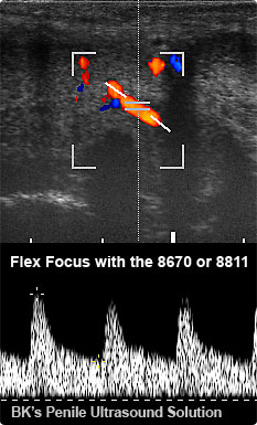
Varicoceles
The presence of varicoceles is a leading factor in the cause of male infertility. Varicoceles can be reliably diagnosed using ultrasound.

The use of color Doppler is a particularly easy technique to evaluate varicoceles. Performing a valsalva maneuver during an ultrasound examination can improve diagnostic confidence.

To perform a varicoceles examination, you begin by scanning the scrotum using the 8670 transducer to make a baseline measurement. The patient is asked to perform a valsalva movement, causing the venous pressure to rise and dilating the veins. The subsequent changes can be examined using ultrasonography. In a patient with varicoceles, it is possible for Doppler ultrasound to show blood flow reverse direction when flowing out of the testicles.

 
Testicular torsion detection
Torsion is a twisting of the spermatic cord resulting in the obstruction of the blood vessels. Color Doppler ultrasound can be useful in the examination and diagnosis. Color Doppler ultrasound can show a decreased or absence of blood flow in the testis. Testicular torsion must be diagnosed urgently. Ultrasound provides a non-invasive, fast, and effective diagnosis.
Testicular ultrasound tools
The 8670 and the Flex Focus scanner is a great choice for Doppler and Testis scanning. The large bandwidth and high maximum scanning frequency of the 8670, provides superb image detail in the near field. A Doppler scan can be used to evaluate the vascular pattern.
 
 
Penile Ultrasound

The 8811 and the 8670 transducers are great choices for Doppler and Penile scanning.

Erectile Dysfunction

Using spectral Doppler it is possible to examine and quantify blood flow. To perform the examination, a vasoactive substance (e.g. prostaglandin) is injected to produce an erection, and if successful, the problem can most likely be corrected. A comparison of arterial velocity before and during erection can many times provide an explanation to the cause of erectile dysfunction.


Penile Doppler
Doppler imaging can be an invaluable tool for the evaluation of impotency.

Erectile dysfunction has various causes: Arterial insufficiency, venous leakage, cavernosal disorders, or can arise from a neurological disorder. During the exam, Doppler is used to assess the arterial velocity before and after injection of a vasoactive substance.


Doppler Ultrasound
A noninvasive diagnostic tool used to evaluate vascularity.

Doppler solutions
The UltraView and the 8818,8808e, 8667, 8823, 8670, 8811, 8848, 8658, or 8820e

Prostate Doppler

with the Prostate Triplane 8818,
Prostate Biplane 8808e,
Endfire Curved Array 8667,
Endocavity Biplane 8848 or 
Endovaginal 8819 transducer.

Color Doppler can help guide prostatis detection and focal lesion biopsies during diagnosis and therapy follow-up procedures.

Kidney Doppler

with the Curved Array 8823 or 8820e transducer.

Color and spectral Doppler help evaluate kidney obstruction, renal artery stenosis and kidney graft control.

Testis Doppler

with the Linear Array 8670 or 8811 transducer.

To help assess varicoceles and testicular inflamation and to aid in the diagnosis of testicular torsion.

Penile Doppler

with the Linear Array 8670 or 8811 transducer.

To help evaluate impotency.


Learn more about the benefits of Doppler for


The largest portfolio of Contrast solutions

With an UltraView solution you have all the tools necessary to locate and map lesions, evaluate blood flow, biopsy suspicious areas, and follow-up after therapy. The UltraView together with our surgical, abdominal, small part, and endorectal transducers, are ideal for Contrast enhanced imaging.

Kidney & Prostate
See Contrast Solutions below for Contrast enhanced imaging of the kidney and prostate.

Urologic Contrast Solutions
UltraView and 8818, 8848, 8823, 8666, or 8820e.


* Contrast Enhanced Ultrasound (CEUS) has not been market cleared by the FDA for use in the USA. Consult your local authorities for CEUS market clearance information in your country.

Contrast Solutions
 
8823

8818

 
Kidney
Contrast for the kidney is particularly useful in the examination of anatomic variations, complex cystic lesions, suspected cystic carcinoma, renal trauma, and residual tumour after RF and other ablative techniques. During follow-up, Contrast may be used in detecting the presence of vascularity after performing an ablative treatment, or for example, monitoring kidney transplants.

Learn about the Guidelines and Good Clinical Practice Recommendations for Contrast Enhanced Ultrasound (CEUS) at EFSUMB.org (European Federation of Societies for Ultrasound in Medicine and Biology).
 

Urologic Ultrasound of the Kidney

Renal ultrasound with the flex Focus and BK Medical transducers gives you:

  • Superior image quality
  • Easy intercostal imaging capabilities

for kidney examination, measurement and biopsy.

Kidney Ultrasound
with the Curved Array 8823 or 8830 Transducer

  • High image resolution at high frequencies
  • Rounded imaging surface, minimizing patient discomfort with easy access between the ribs and better contact with the skin
  • Range of single-use and reusable needle guides for interventional procedures


Prostate
Contrast can be used on the prostate. The 8818 and 8848 transducers are ideal to measure vascularity after ablation to verify success.


* Contrast Enhanced Ultrasound (CEUS) has not been market cleared by the FDA for use in the USA.

Our newest high resolution 3D endocavity transducer - the 3DART 8838 - gives you such detailed images that you can:
  • See the entire gland geometry and size
  • Clearly identify prostate borders
  • Differentiate all prostate zones

To help you make more confident diagnostic decisions and plan treatment faster.

HistoScanning™ workstation

Pro Focus UltraView and the 8818 tranducer.

3D solutions

UltraView or Flex Focus scanner.
Transducer
8808, 8818, 8848, or 8658 .
A magnetic wheel mover with professional 3D software,

or freehand.

3D Ultrasound Imaging Solutions

3D ultrasonography is available on a wide range of BK transducers as a standard option, and several of our urology transducers offer tracked 3D imaging capabilities.

How 3D imaging works

3D Ultrasound of the Prostate

  • Gives you a simultaneous sagittal, transverse and coronal view of the prostate to detect possible extra-capsular tumor extension
  • Provides valuable information to plan for alternative therapies, like robotic prostatectomy and radioactive treatment of the prostate
  • May make it easier to determine whether the “prostate capsule” has been penetrated, a key factor in staging
  • Gives access to a new, wider range of viewing planes that are unavailable with 2D imaging

With 3D ultrasound, images can be manipulated and rotated to get the best possible view.

After these images have been saved, they can be reviewed as many times as needed. Examination and potentially diagnosis can then be achieved offline when it is most convenient for the operator and patient, leading to faster patient turnaround.


The advantages and benefits of 3D imaging.

  Benefits of 3D prostate imaging compared to 2D:
  • Improves staging accuracy compared to 2D, which lacks in-depth information.
  • Gives access to a new, wider range of viewing planes that are unavailable with 2D scanning.
 
  Advantages of working with 3D:
  • Easily distinction between prostate zones and see small lesions.
  • Images can be manipulated and rotated to get the best possible view, and these images can be saved and reviewed as many times as needed.
  • A diagnosis can be achieved offline when it is most convenient for the operator and patient, leading to faster patient turnaround.

The role of 3D in prostate diagnostics.

3D imaging of the prostate


With 3D imaging, spatial relationships much clearer, so urologists can better assess the extent of disease. It provides valuable information to plan for alternative therapies, for example, external beam radiation or prostatectomy/radioactive seed implantation. In particular, it may be easier to determine whether the “prostate capsule” has been penetrated, a key factor in staging.

3D imaging of the prostate enables a simultaneous view of the sagittal, transverse and, of particular importance, the posterior-anterior coronal view of the gland to detect possible extra-capsular tumour extension.

HistoScanning™ workstation

Pro Focus UltraView and the 8818 or 8848 tranducers

HistoScanning™ compatible products for the prostate


HistoScanning™ is an affordable, non-invasive diagnostic imaging tool for prostate diagnostics and is offered only on BK solutions. This exciting, revolutionary technology promises to change the way specialists perform prostate diagnosis and biopsies. HistoScanning™ may help physicians make more informed treatment decisions for their patients.

To get started, you will need the licensed HistoScanning-ready UltraView scanner and to license the HistoScanning workstation from AMD. The system is compatible with the 8818 or 8848 transducer and requires a magnetic wheel mover for accurate motorised sagittal and/or transverse 3D prostate scanning.

Prostate HistoScanning™ is a proprietary tissue characterization technology developed to differentiate, characterize and visualize prostate tissue, based on the analysis of backscattered ultrasound.

A Unique Ultrasound Application to Aid:

  • Treatment selection
  • Individual treatment planning
  • Triaging patients for active surveillance

HistoScanning is compatible with our Prostate Triplane Transducer 8818 and magnetic wheel mover for accurate motorized sagittal 3D prostate scanning.

To get started, you need:

  • Licensed HistoScanning-ready UltraView system
  • HistoScanning workstation from Advanced Medical Diagnostics (AMD)

HistoScanning is CE marked. HistoScanning has not been market cleared by the FDA for sale in the USA.