|
| |
Ultrasound Equipment
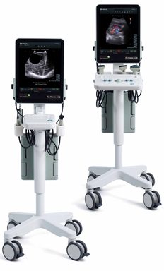
Pro Focus 2202 Ultraview:
Making You Confident with Ultrasound
Pro Focus Ultraview is a full featured ultrasound
system, providing premium images that make a diagnostic difference.
The high performance Ultraview system offers IQPACTM
technology, contrast imaging, HistoScanningTM interface and a complete
range of innovative, dedicated transducers designed to meet the clinical
needs of specialists.
The compact mobile design brings ultrasound right to
the point of care. With features such as adjustable settings and user
friendly navigation, Ultraview is designed to work with you.
Developed as an integrated clinical tool, the
Ultraview streamlines workflow and provides compatibility with products
needed for confident diagnostics and choosing the best course of patient
treatment.
A
diagnostic toolkit
With a Pro
Focus Ultraview solution you have all the tools necessary to locate and
map lesions, evaluate blood flow and biopsy suspicious areas. Improved
workflow makes it easy to create clear images for an accurate diagnosis.
our transducers cover all major specialities and both near field and far
filed scanning
Monitor
therapeutic intervention
With the
Ultraview system, you can monitor breast biopsy, cyst drainage,
radioactive seed implant, or other interventional therapies including
RFA, cryo, microwave and laser. the ultraview helps you guide the tip of
the catheter during RF ablation and drainage.
A measure of your success
The
Ultraview is compatible with a range of transducers capable of
contrast-enhanced ultrasound examinations. Contrast imaging may be used
in detecting the presence of vascularity after perfoming an ablative
treatment.
Work confidently and efficiently with
ultrasound
The Pro Focus optimizes your workflow by providing you with the
information you need quickly, so you can make confident decisions. With
features like an intuitive and easy-to-use interface and automatic
optimization of transducer and application settings, the Pro Focus is
designed to provide high-quality images immediately.
Ultrasound made simple
Features:
- Intuitive and easy-to-use interface
- Seamlessly integrated 3D
- Quick and simple image capture
- Contrast harmonic imaging
The Pro Focus provides a choice of over 20 versatile
transducers and puncture guides to suit a whole range of examinations.
Switch transducer and the Pro Focus instantly adapts to the new
situation.
An easy-to-use internal archiving system allows you to
save and replay images, video clips, 3D data sets and reports on the Pro
Focus hard drive. Fully integrated DICOM* capabilities, built-in CD- ROM
drive and USB ports means that images from the Pro Focus can be quickly
transferred to a standard computer, from which they can be e-mailed to
colleagues for second opinions, for example.
Note: Some of the features mentioned above are available as options.
Transducers recommended for the
Pro Focus 2202
*DICOM is the registered trademark of the National
Electrical Manufacturers Association for its standards publications
relating to digital communications of medical information. |
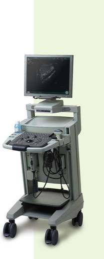
The Pro Focus 2202 Ultraview Ultrasound System, together with specialized
transducers, provides an unbeatable package. It makes it easy for you to
access the information you need; it’s straightforward to operate and has
excellent image quality. |
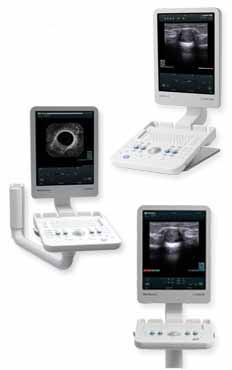
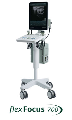
Times have changed, and so has mobile
ultrasound
The image quality on the Flex Focus is like nothing you
have ever seen on a mobile system.
The Flex Focus features:
- IQPAC™ technology for speckle reduction and
better organ definition. IQPAC technology optimizes ultrasound
images through Enhanced Tissue Definition (ETD) and Angular
Compound Imaging
- (ACI). Reducing ultrasound speckle enhances the
anatomically correct continuous borders of organs and improves the
ability to clearly visualize margins of lesions.
The Flex Focus and IQPAC technology because Image is
Everything.
|
 |
|
|
|
|
| |
|
|
|
|
|
|
|
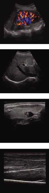
Take your practice with you anywhere. The Flex
Focus is lightweight, small, goes with you to the
point-of-care. |
|
|
A good overall traveller
Imagine what you can do. Flex
Focus loves to go wherever you go. On the wall, on your desk,
in your car and even on an airplane. With the
Flex Focus, take your practice with you anywhere.
|
UROLOGY
|
|
 |
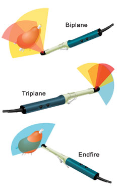
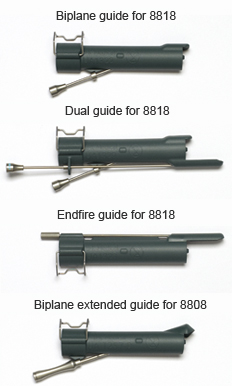
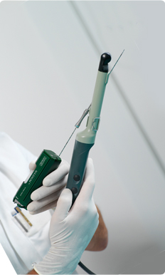
Ultrasound Transducer, SeeDOS code : 8818
Triplane – all prostate zones with one
transducer
- unsurpassed images in
3 visionary planes
- switch between
prostate zones at the touch of a button
- increase diagnostic
value with 3D, Contrast and Doppler
Easy and comfortable to use
- take confident apical
biopsies with endfire array
- biopsy the
peripheral, transition and central zones with
simultaneous biplane
- one-time insertion
and minimal manipulation using disposable dual guide
Read more about our sterile single-use
needle guides
here.
8818 Applications
- Transrectal prostate
scanning
- Transrectal puncture
and biopsy
- Transperineal
puncture and biopsy
- Transvaginal scanning
- Spectral and CFM
Doppler examinations
- Tissue harmonic
imaging
- Contrast imaging*
|
|
|
|
|
|
|
|
|
|
|
Conventional examinations are not
able to provide the details needed
to understand:
|
|
|
| |
|
|
|
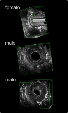
Normal
Anatomy
Normal anorectal anatomy of
men and women is different
in a few important ways. While the length of the
internal
sphincter is the same, understanding the differences is
crucial to being able to understand normal anatomy and
identify external sphincter tears in women.
See more ultrasound image
examples of normal anatomy for
both men and women.
|
|
|
|
|
|
|
Anal Tears
Anal tears are mainly the result of an obstetric or
iatrogenic injury. They are found either in the external
anal sphincter, internal anal sphincter or a combination
of both.
On the ultrasound image scarring is
either hypo- or hyperechoic. Internal anal sphincter
tears are breaks in the normal hypoechogenic ring. Once
a tear is identified, it needs to be classified
according to the radial and longitudinal extension.
|
|
|
|
Fistulas
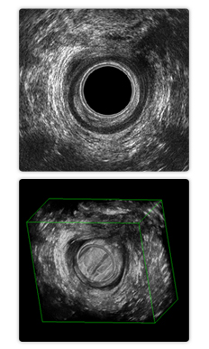
3D imaging of fistulas offers a
significant advantage over conventional 2D ultrasound.
Identifying the fistulas internal opening and following
its tracts are nearly impossible with 2D imaging.
A high resolution 3D data acquisition
takes approx. 30 seconds with the 2052 transducer. If an
external opening is identified, some doctors introduce
hydrogen peroxide H2O2
(3–5%) into the opening immediately before acquiring a
3D data set. During this short period, the H2O2 enhances
the fistula tracts so that they appear as bright white
structures on the ultrasound image. Aerated and diluted
lidocaine gel may also be introduced in
an external opening as an ultrasound
enhancing medium
|
|
|
|
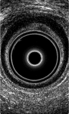
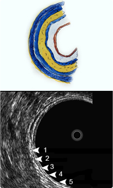
Rectal Cancer
Ultrasound studies show all layers of
the rectal wall, represented in the image as 4
hyperechoic and 3 hypoechoic structures
(assuming that the layer line between the
inner circular and the other longitudinal layers are
seen in the muscularis propria).
In the anatomical representation of the
recturm on the left ,
the layers represent:
| 1 |
the hyperechoic
interface between the waterfilled balloon and
the mucosa |
| 2 |
the hypoechoic
deep mucosa (lamina propria and muscularis
mucosae) |
| 3 |
the hyperechoic
submucosa |
| 4 |
the hypoechoic
muscularis propria (in some cases seen as 2
layers: inner circular and outer longitudinal
layer) |
| 5 |
the hyperechoic
interface between the rectal wall and the
perirectal fat tissue |
|
|
|
|
Ultrasound Transducer, SeeDOS Code 8818
|
|
| Frequency Range |
4 - 12 MHz |
Contact surface
(acoustic) |
34.4 x 5.5 mm |
| Focal range |
3 - 60 mm |
| Scanning modes |
B, M,BCFM, Doppler,
Contrast*, Tissue Harmonic |
| Frame Rate |
60 Hz |
| Image field(expanded) |
Triplane / 140° |
| Dimensions |
36 x 39 x 323 mm |
| Weight |
230 g |
| Disinfection |
Immersion,
STERIS SYSTEM 1**
STERIS SYSTEM 1E,
STERRAD 50,100S and 200 |
|
|
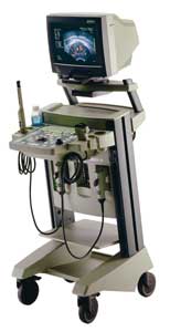
Falcon 2101 EXL
Benefits |
 |
-
integrated
image storage as standard
-
DICOM-compatible
output
-
high capacity
image review
-
ECG option
-
large
diameter trackball
-
moveable
multifocus on all electronic transducers
-
customizable
keyboard and measurements
-
B-K Medical's
Instant Recall feature
-
Palm Control
Unit (PCU) for remote use
-
multitude of
uses with an imaging bandwidth of 1-15 MHz
-
highest
resolution in its class
-
sleek,
compact design
-
extensive
measurement and calculation facilities
-
simultaneous
split screen feature
-
optional
integrated 3D package
-
simple and
convenient operator interface
-
fully
adjustable monitor and front panel
|
|
Type 8658 Ultrasound Probe
|
An ideal choice for use in the treatment of prostate diseases
For more details of this ultrasound probe please click here | |
Type 8658/8558 transducer is an elegantly shaped instrument with multifrequency, biplane and colour Doppler capabilities. It is an excellent choice for all transperineal procedures including brachytherapy because of its unique long sagittal array for a base to apex view.
The ability to scan both transversely and sagittally also makes prostate volume determination particularly easy when using the HWL method – even more so when used in combination with the scanner’s split-screen facility.
| |
Type 8658/8558 Benefits
|
 |
slender design for patient comfort |
|
handle-based controls |
|
|
easy to attach needle guide for transperineal treatment methods |
|
|
compatible with all leading manufacturer’s stepper and template units for ultrasonically guided seed implantation procedures (brachytherapy) - Go To Stepper/Stabilizer/Template |
|
|
Multifrequency imaging: Type 8658/8558 scans at 5.0, 6.5, and 7.5 MHz.
|
Type 8658 is for use with our 2102 XDI, 2102 and 2101 scanners.
Type 8558 is for use with our 2001, 2002, 2002 ADI, 2003 and 3101 scanners.
| |
Images
Please click here for an image of a transverse section of the prostate with visualisation of the peripheral zone using the type 8558 probe.
|
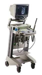
HAWK 2102 EXL:
Flexible ultrasound in a highly integrated multipurpose colour scanner
Available with type
8658 ultrasound probe for use in the prostate seed implant treatment
technique with additional doppler functions.
The high-definition Hawk 2102 EXL gives you an ideal
combination of high performance, versatility and ease of use, making it
suitable for a wide variety of applications.
It offers flexibility on many levels, from its
user-definable features to its comprehensive choice of specially
designed B-K Medical transducers with corresponding puncture and biopsy
equipment.
The Hawk includes a full range of Doppler functions.
It also features tissue harmonic imaging with True Echo Harmonics™ to
help you image difficult to scan patients. |
The Hawk 2102 EXL colour scanner offers high performance and great
versatility.
|
|
Highlights of the Hawk 2102 EXL
include:
- Frequency range of 1-20 MHz provides the
highest resolution in its class.
- Four transducer connectors (three array
and one mechanical).
- B-K Medical's patented Continuous Uniform
Focusing technology ensures optimal resolution at all image depths.
- Compact and mobile, so it can be used
wherever and whenever you need it.
- Ergonomic, with height-adjustable keyboard
and monitor and logically grouped controls.
- Full Doppler functionality, including CFM,
Power Doppler, CW (not cleared for use in USA), and duplex and
triplex imaging with real-time calculations.
-
True Echo Harmonics™ gives improved
imaging of difficult to scan patients.
- Full 15" flicker-free monitor provides
optimum viewing.
- Optional fully integrated 3-D package,
including tracked pullback support for relevant transducers.
- Simultaneous split screen feature lets you
display two active ultrasound images on the monitor at the same
time.
- Optional fully integrated DICOM® package*.
- Convenient storage space on the unit
itself, with a compartment under the keyboard for accessories.
- Upgradability ensures you of long-tem
investment security.
Transducers recommended for
the Hawk 2102 EXL
DICOM Conformance Statement (PDF) available on
request.
*DICOM is the registered trademark of the National
Electrical Manufacturers Association for its standards publications
relating to digital communications of medical information. |
If you are interested in any of the products mentioned here, please complete a
Quotation Request Form or an
Enquiry Form so that we may promptly respond to your detailed request. |
TOP OF PAGE
Please contact Colin Walters at cwalters@seedos.com if you would like further information or you have questions
or comments about this web site.
SeeDOS Ltd, 26, The Maltings, Leighton Buzzard, Bedfordshire LU7 4BS, United Kingdom
Tel: +44 1525 850 670 • Fax: +44 1525 850 685
|
|